Table of Contents >> Show >> Hide
- What Is Cutaneous Mastocytosis?
- Main Types of Cutaneous Mastocytosis
- Cutaneous Mastocytosis Symptoms
- What Causes Cutaneous Mastocytosis?
- Common Triggers That May Worsen Symptoms
- How Cutaneous Mastocytosis Is Diagnosed
- Treatment for Cutaneous Mastocytosis
- Living With Cutaneous Mastocytosis
- When to Call a Doctor
- Outlook and Prognosis
- Experiences and Practical Lessons From Living With Cutaneous Mastocytosis
- Conclusion
Cutaneous mastocytosis is one of those medical terms that sounds like it belongs in a sci-fi lab, but it actually describes a rare skin condition involving mast cells. Mast cells are immune cells that normally help protect the body. They sit in tissues, including the skin, and release chemical messengers such as histamine when the immune system senses trouble. In cutaneous mastocytosis, too many mast cells collect in the skin, where they can cause spots, itching, flushing, swelling, and sometimes blisters.
The condition is most often seen in infants and young children, although adults can develop it too. In many children, cutaneous mastocytosis improves over time and may fade by adolescence. In adults, doctors often take a closer look to make sure the condition is limited to the skin and not part of systemic mastocytosis, which can involve bone marrow or internal organs.
The good news? Many cases are manageable. The less-fun news? Mast cells can be dramatic little divas. Heat, friction, certain medicines, stress, infections, or even vigorous rubbing of the skin may trigger symptoms. Understanding the signs, causes, diagnosis, and treatment options can help patients and families feel less confused and more prepared.
What Is Cutaneous Mastocytosis?
Cutaneous mastocytosis is a form of mastocytosis that primarily affects the skin. The word “cutaneous” means skin-related, while “mastocytosis” refers to an abnormal increase or buildup of mast cells. These cells are part of the immune system and are loaded with substances that can cause allergy-like reactions when released.
When mast cells are activated, they may release histamine and other mediators. This release can lead to itching, redness, hives, swelling, flushing, and irritation. Think of mast cells as tiny security guards. In most people, they sound the alarm only when needed. In cutaneous mastocytosis, there are too many guards in one area, and some of them are a little too eager to press the alarm button.
Main Types of Cutaneous Mastocytosis
Maculopapular Cutaneous Mastocytosis
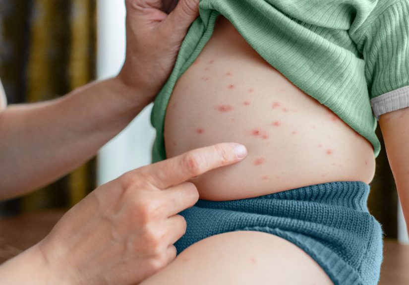
Maculopapular cutaneous mastocytosis, historically called urticaria pigmentosa, is the most common form. It usually appears as multiple brown, tan, reddish-brown, or pinkish spots or bumps on the skin. These spots may become raised, itchy, or swollen when rubbed or exposed to heat.
Solitary Cutaneous Mastocytoma
A mastocytoma is usually a single patch, plaque, or nodule made up of mast cells. It often appears in infancy or early childhood. The area may blister, swell, or become itchy when irritated. Many solitary mastocytomas improve naturally as a child grows.
Diffuse Cutaneous Mastocytosis
Diffuse cutaneous mastocytosis is rarer and can involve widespread thickening, redness, or blistering of the skin. Because the skin involvement may be more extensive, children with this form may need closer monitoring by dermatology, allergy, or pediatric specialists.
Cutaneous Mastocytosis Symptoms
Symptoms can vary widely. Some people have only a few spots and mild itching. Others may have more noticeable reactions, especially when the skin is rubbed, heated, or irritated.
Common Skin Symptoms
- Brown, red-brown, tan, pink, or yellowish spots or bumps
- Itching, especially after heat, friction, or sweating
- Skin swelling or hive-like reactions
- Flushing or warmth of the skin
- Blistering, especially in infants or young children
- Thickened or leathery skin in rare diffuse cases
Darier Sign
One classic clue is called Darier sign. This happens when rubbing or stroking a lesion causes it to become red, swollen, itchy, or hive-like. It is not a party trick to perform at home repeatedly, because irritating the skin can make symptoms worse. Doctors may check for it carefully during evaluation.
Body-Wide Symptoms Can Happen
Although cutaneous mastocytosis mainly affects the skin, mast cell mediator release can sometimes cause symptoms beyond the rash. These may include flushing, stomach cramps, diarrhea, nausea, headache, lightheadedness, or rapid heartbeat. Severe allergic-type reactions are uncommon but possible, especially in people with extensive disease or strong triggers.
What Causes Cutaneous Mastocytosis?
Cutaneous mastocytosis is commonly linked to changes in the KIT gene, which helps regulate mast cell growth and survival. Many cases involve somatic genetic changes, meaning the change occurs in certain cells and is not inherited from a parent. Rarely, mastocytosis can appear in more than one family member.
Importantly, cutaneous mastocytosis is not contagious. You cannot catch it from someone else, and it is not caused by poor hygiene, diet mistakes, or a mysterious curse from a laundry detergent commercial. The condition involves abnormal mast cell accumulation and activation, not an infection.
Common Triggers That May Worsen Symptoms
Many people with cutaneous mastocytosis notice symptoms flare after specific triggers. Triggers are not the same for everyone, so keeping a symptom diary can be surprisingly helpful.
- Heat, hot baths, or sudden temperature changes
- Friction from tight clothing, scratching, or rubbing
- Vigorous exercise or overheating
- Stress or strong emotions
- Infections or fever
- Alcohol in adults
- Certain medications, depending on individual sensitivity
- Insect stings or bites
- Some foods in sensitive individuals, though food is not always a trigger
Trigger management does not mean living inside a bubble. It means learning which situations make symptoms worse and planning around them. For example, a child who flares after hot baths may do better with lukewarm water, soft towels, and gentle skin care.
How Cutaneous Mastocytosis Is Diagnosed
Diagnosis often starts with a careful skin exam and medical history. A doctor will ask when the spots appeared, whether they itch or swell, what triggers symptoms, whether there are digestive or allergy-like symptoms, and whether there is a history of severe reactions.
Skin Examination
A dermatologist may recognize the pattern of lesions, especially if spots become swollen or red after gentle irritation. The appearance, distribution, age of onset, and symptom pattern all matter.
Skin Biopsy
A skin biopsy may be used to confirm the diagnosis. During a biopsy, a small sample of affected skin is examined under a microscope. Special stains can show increased mast cells in the skin.
Blood Tests
Doctors may order blood tests, including serum tryptase. Tryptase is an enzyme released by mast cells, and higher levels can suggest a larger mast cell burden or systemic involvement. Mild changes can occur, especially in children with extensive skin lesions, so results must be interpreted by a clinician familiar with mast cell disorders.
Checking for Systemic Disease
In children with typical cutaneous mastocytosis and no concerning symptoms, extensive testing may not always be needed. In adults, persistent skin lesions may prompt additional evaluation because adult-onset mastocytosis is more often associated with systemic disease. Depending on the case, doctors may consider additional blood work, genetic testing, imaging, or bone marrow evaluation.
Treatment for Cutaneous Mastocytosis
Treatment depends on age, symptoms, severity, and whether the condition is limited to the skin. Many children need only reassurance, trigger avoidance, and symptom relief when flares occur. Adults and people with frequent or severe symptoms may need a more structured treatment plan.
Trigger Avoidance
The first treatment step is often practical: reduce known triggers. That may mean avoiding hot showers, choosing loose cotton clothing, using gentle fragrance-free skin products, preventing overheating, and treating fevers promptly. It is simple advice, but simple does not mean useless. Sometimes the skin just wants fewer dramatic plot twists.
Antihistamines
H1 antihistamines may help reduce itching, hives, flushing, and skin irritation. H2 blockers may be used when stomach symptoms such as acid discomfort, cramping, or nausea are part of the picture. A clinician can recommend the best option and dose based on age and symptoms.
Topical Corticosteroids
Short courses of topical corticosteroid creams may be prescribed for inflamed or itchy lesions. These should be used exactly as directed, because overuse can thin or irritate the skin.
Mast Cell Stabilizers
In selected cases, doctors may use mast cell stabilizers such as cromolyn sodium, especially when gastrointestinal symptoms are present. This treatment is not for everyone, but it can help certain patients reduce mediator-related symptoms.
Epinephrine Autoinjector
Some patients may be advised to carry an epinephrine autoinjector, especially if they have had severe reactions or are considered at higher risk for anaphylaxis. Families should be taught when and how to use it. Emergency planning can feel scary at first, but it is really about being prepared, not panicked.
Phototherapy
Adults with troublesome skin lesions may sometimes be treated with phototherapy, such as ultraviolet-based treatment under medical supervision. Phototherapy is not a casual tanning-bed situation; it is a controlled medical treatment with risks and benefits that should be discussed with a specialist.
Treatment of Systemic Mastocytosis
If evaluation shows systemic mastocytosis, treatment may involve additional therapies and specialist care. This can include symptom-control medicines, monitoring, and in certain cases targeted treatments. Cutaneous mastocytosis and systemic mastocytosis overlap in name, but the management plan can be very different.
Living With Cutaneous Mastocytosis
Daily life with cutaneous mastocytosis is often about learning patterns. A parent may notice that a toddler’s spots swell after a hot bath. An adult may realize that stress, alcohol, or overheating makes flushing worse. These observations are useful because they turn a mysterious condition into something more predictable.
Skin care should be gentle and boringin the best possible way. Fragrance-free cleansers, moisturizers, lukewarm water, soft fabrics, and sun protection can help reduce irritation. Scrubbing the skin aggressively is usually a bad idea. The goal is calm skin, not skin that feels like it has survived a car wash.
When to Call a Doctor
Medical attention is important if symptoms are worsening, lesions are blistering frequently, a child seems unwell, or there are body-wide symptoms such as faintness, breathing trouble, severe abdominal pain, repeated vomiting, or signs of anaphylaxis. Adults with new mastocytosis-like skin lesions should also be evaluated, because adult-onset disease deserves careful assessment for systemic involvement.
Patients should also talk to a healthcare professional before surgery, anesthesia, or starting new medications, especially if they have a history of severe mast cell reactions. A written action plan can be helpful for schools, caregivers, travel, and emergency settings.
Outlook and Prognosis
The outlook depends on the type of cutaneous mastocytosis, age of onset, and whether there is systemic involvement. In many children, especially those with skin-limited disease, symptoms improve over time and may fade significantly by puberty. Solitary mastocytomas often improve on their own.
Adults may have a more persistent course and may require ongoing monitoring. The key is accurate diagnosis, symptom control, trigger awareness, and follow-up with clinicians who understand mast cell disorders.
Experiences and Practical Lessons From Living With Cutaneous Mastocytosis
Although every case is different, people living with cutaneous mastocytosis often describe a similar emotional journey: confusion first, then detective work, then confidence. At the beginning, the spots may look like bug bites, eczema, birthmarks, hives, or a rash that refuses to follow the usual rash rules. Families may bounce between creams, laundry changes, and internet searches before a dermatologist finally names the condition.
One common experience is learning that the skin can react quickly. A child may look perfectly comfortable in the morning, then develop red, raised patches after playing outside on a warm day. An adult may notice flushing after a hot shower or itching after wearing tight exercise clothing. These reactions can be frustrating, but they also provide clues. Once patterns are recognized, small adjustments can make a big difference.
For parents, bath time is often an early lesson. Hot water may trigger itching or swelling, so lukewarm baths become the new normal. Towels are used for patting rather than rubbing. Moisturizer becomes part of the routine. The bathroom may start to look like a tiny dermatology spa, minus the cucumber water and dramatic music.
School and daycare planning can also matter. Teachers and caregivers may need a simple explanation: the spots are not contagious, scratching or rough play can irritate them, and certain symptoms should be reported. If an epinephrine autoinjector is prescribed, adults caring for the child should know where it is and how to use it. Clear instructions reduce fear and prevent overreaction.
Adults with cutaneous mastocytosis may face a different challenge: uncertainty. Because adult-onset mastocytosis can sometimes be associated with systemic disease, testing and follow-up may feel intimidating. Many patients find it helpful to write down symptoms before appointments, including flushing, itching, digestive symptoms, fatigue, medication reactions, and possible triggers. A well-kept symptom diary can turn a rushed appointment into a more productive conversation.
Another practical lesson is that “avoid triggers” should be realistic. Nobody can avoid heat, stress, infections, exercise, and friction forever. Life includes weather, deadlines, waistbands, and occasional chaos. The goal is not perfection. The goal is reducing avoidable flares and knowing what to do when symptoms appear.
Support can be valuable too. Rare conditions can feel isolating because friends may not understand why a skin condition needs an action plan. Connecting with reputable patient organizations, specialists, and informed healthcare teams can help patients feel less alone. Good education replaces panic with preparation.
Perhaps the most encouraging experience many families report is that childhood cutaneous mastocytosis often becomes easier with time. Spots may fade, reactions may decrease, and routines become second nature. For adults, the path may involve more monitoring, but symptom control is still possible. With the right care plan, cutaneous mastocytosis can become something a person managesnot something that manages them.
Conclusion
Cutaneous mastocytosis is a rare skin condition caused by an abnormal buildup of mast cells in the skin. It can cause brown or red-brown spots, itching, flushing, swelling, and sometimes blistering. While the condition can look alarming, especially in children, many cases are manageable and may improve over time.
Diagnosis usually involves a skin exam, medical history, and sometimes a biopsy or blood testing. Treatment focuses on avoiding triggers, calming symptoms, using antihistamines or topical medicines when needed, and preparing for rare severe reactions. The best plan is individualized, because mast cells are not exactly famous for being predictable.
Anyone with suspected cutaneous mastocytosis should work with a qualified healthcare professional, especially if symptoms are severe, new in adulthood, or associated with body-wide reactions. With the right information and care, patients and families can move from “What is happening to my skin?” to “We know what this is, and we have a plan.”