Table of Contents >> Show >> Hide
- Step 1: Confirm it’s vertigo (spinning) vs. other kinds of dizziness
- Step 2: Screen for emergency “red flags” before you do anything else
- Step 3: Use the “timing + triggers” method (it beats guessing)
- Step 4: Build a “vertigo snapshot” your clinician can actually use
- Step 5: Check for ear clues (because your inner ear is dramatic)
- Step 6: Look for migraine features (even if you don’t always get head pain)
- Step 7: Ask: “Was there an infection or sudden ‘one bad week’?”
- Step 8: Review medications and substances (the sneaky culprits)
- Step 9: Check blood pressure patterns, especially orthostatic changes
- Step 10: The exam that matters most: eyes + balance + neurologic checks
- Step 11: If symptoms are position-triggered, your clinician may do the Dix-Hallpike test
- Step 12: Vestibular and hearing tests may be ordered to pinpoint the cause
- Step 13: Decide when labs or imaging are (and aren’t) useful
- Putting it together: a fast “diagnosis-ready” checklist
- Real-World Experiences: What Diagnosing Vertigo Often Feels Like (and What Helps)
- Conclusion
Vertigo is not just “feeling dizzy.” It’s that unmistakable, unfair amusement-park ride sensationexcept you didn’t buy a ticket, and the ride operator is your inner ear. The tricky part? Vertigo is a symptom, not a diagnosis. So the real question is: what’s causing itand how do you figure that out quickly and safely?
This guide walks through the same logic clinicians use to diagnose vertigotranslated into normal-human language, with a little humor and a lot of clarity. You’ll learn how to describe symptoms in a way that helps your provider narrow the cause, what tests they may use, and when vertigo is a “call-now” emergency.
Important: This article is educational and not a substitute for medical care. If you have severe symptoms or “red flags,” seek urgent help.
Step 1: Confirm it’s vertigo (spinning) vs. other kinds of dizziness
People say “dizzy” to mean at least four different things: lightheaded (like you might faint), off-balance (like you’re walking on a boat), woozy/spacey, or true vertigo (spinning/motion that isn’t happening). Diagnosis starts by sorting these out.
What vertigo feels like
- You feel like the room is spinning, tilting, or moving.
- You feel like you are spinning or being pulled to one side.
- It may come with nausea, sweating, or trouble walking straight.
Why this matters: different sensations point to different systemsinner ear, brain, heart/blood pressure, medications, anxiety, or vision. Calling everything “vertigo” is like telling a mechanic your car makes “a noise.” True, but we can do better.
Step 2: Screen for emergency “red flags” before you do anything else
Some causes of vertigo are annoying but benign. Others can be seriousespecially if vertigo is sudden and severe or paired with neurologic symptoms. Use this as a safety check, not a self-diagnosis contest.
Call emergency services or go to the ER now if vertigo comes with:
- Weakness, numbness, facial droop, or trouble speaking
- New double vision, severe trouble walking, or inability to stay upright
- Worst headache of your life, neck stiffness, or fainting
- Chest pain, severe shortness of breath, or a new irregular heartbeat sensation
- Sudden hearing loss (same-day urgent evaluation is recommended)
Translation: if your brain seems involvedor you can’t safely standthis is not the moment for a “let’s see if it passes” approach.
Step 3: Use the “timing + triggers” method (it beats guessing)
Clinicians often diagnose vertigo by pattern recognition. The pattern comes from two things: timing (how long episodes last) and triggers (what sets it off).
Common patterns that help narrow the cause
- Triggered, brief episodes (seconds to a minute) when you roll over in bed, look up, or bend down: often points toward BPPV (benign paroxysmal positional vertigo).
- Spontaneous episodes (minutes to hours) that come and go without a specific head movement trigger: can suggest vestibular migraine or Ménière’s disease, among others.
- Continuous vertigo (hours to days) with nausea and imbalance: may fit vestibular neuritis (inner ear nerve inflammation) or, less commonly, a central cause that needs urgent evaluation.
This step is a big deal because it helps your provider choose the right exam maneuvers and decide whether testing (or imaging) is needed.
Step 4: Build a “vertigo snapshot” your clinician can actually use
Your appointment will go better if you bring specifics. Think of it like giving your clinician a trailer instead of making them watch the entire movie in chronological order.
Write down (or track in your notes app):
- Start date and whether onset was sudden or gradual
- Episode duration: seconds, minutes, hours, or days
- Triggers: rolling in bed, standing up, visual motion (scrolling), stress, certain foods, loud sounds
- Associated symptoms: hearing changes, ringing (tinnitus), ear fullness, headache, light sensitivity, nausea, palpitations
- Frequency: once, daily, weekly, random chaos
- What helps: sitting still, hydration, sleep, darkness, specific positions
Bonus points (optional but helpful): If you notice your eyes “jumping” during an episode, try recording a short video of your eyes while you look straight ahead. Abnormal eye movements (nystagmus) can be a major cluebut don’t force positions that make you feel unsafe.
Step 5: Check for ear clues (because your inner ear is dramatic)
Many vertigo cases are “peripheral,” meaning they involve the inner ear balance organs or the vestibular nerve. Ear symptoms can strongly shape the diagnosis.
Ear-related clues include:
- Hearing loss (especially fluctuating or one-sided)
- Tinnitus (ringing, roaring, buzzing)
- Ear fullness/pressure
A classic example: recurrent vertigo episodes plus hearing symptoms can suggest Ménière’s disease. Another example: vertigo after a cold with new hearing changes can raise concern for labyrinthitis rather than vestibular neuritis.
Step 6: Look for migraine features (even if you don’t always get head pain)
Vestibular migraine is a common and under-recognized cause of vertigo. And yessome people get the dizziness/vertigo without a classic pounding headache. Rude, but real.
Clues that point toward vestibular migraine
- History of migraines (past or present), motion sickness, or strong family history
- Vertigo episodes linked to stress, sleep disruption, hormonal shifts, or certain foods
- Light/sound sensitivity, visual aura, “brain fog,” nausea during episodes
- Normal ear exam and no consistent hearing loss pattern
Diagnosis is typically clinicalmeaning it’s based on symptoms and patternafter ruling out other causes that look similar.
Step 7: Ask: “Was there an infection or sudden ‘one bad week’?”
If vertigo hits hard, lasts for hours to days, and comes with nausea and imbalance, clinicians consider causes like vestibular neuritis (vestibular nerve inflammation) and labyrinthitis (inner ear inflammation that can affect hearing).
Common story patterns
- Recent viral illness (cold/flu-like symptoms)
- Sudden onset of severe spinning, difficulty walking, nausea/vomiting
- Symptoms gradually improve over days, but balance may feel “off” longer
This is also where safety matters: continuous, severe vertigo can mimic serious central causes. Clinicians use targeted bedside exams to decide who needs urgent imaging and stroke evaluation.
Step 8: Review medications and substances (the sneaky culprits)
Medications can contribute to dizziness, imbalance, and sometimes vertigo-like symptomsespecially when doses change or multiple sedating meds overlap. Bring your full list, including supplements and “only when I can’t sleep” stuff.
Examples of common contributors
- Blood pressure medications (especially if they cause low BP)
- Sleep aids, anti-anxiety meds, muscle relaxers
- Some antidepressants (dose changes can be a factor)
- Alcohol, cannabis, or other substances
- Rarely, certain antibiotics or chemo agents can affect inner ear function
This step doesn’t mean “your symptoms are in your head.” It means your clinician wants to remove avoidable contributors before ordering a parade of tests.
Step 9: Check blood pressure patterns, especially orthostatic changes
Not all “dizzy” episodes are vertigo. If symptoms happen when standing upespecially with dimming vision, weakness, or near-faintingclinicians often check for orthostatic hypotension (a significant BP drop when standing).
What your clinician may do
- Measure blood pressure and pulse lying down, sitting, and standing
- Ask about hydration, recent illness, heat exposure, and salt intake
- Review meds that lower BP
This step helps separate spinning vertigo from lightheadedness/presyncopewhich have very different causes and treatments.
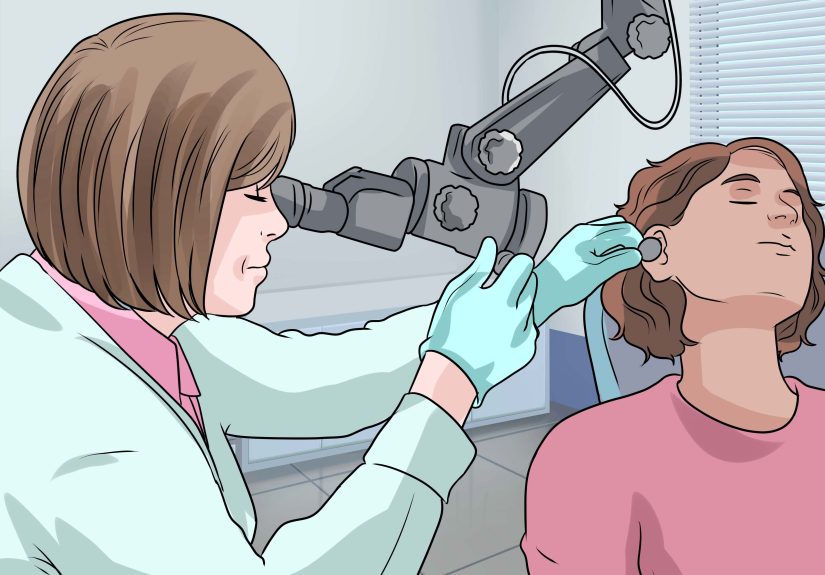
Step 10: The exam that matters most: eyes + balance + neurologic checks
If vertigo had a “tell,” it would be the eyes. The brain and inner ear communicate through reflexes that stabilize your gaze. When that system is disrupted, clinicians often see nystagmus (involuntary rhythmic eye movements) and specific balance findings.
Typical components of the office/urgent care exam
- Eye movement assessment (looking for nystagmus patterns)
- Gait and balance testing (can you walk steadily?)
- Full neurologic screen (strength, sensation, coordination, speech)
- Ear exam and sometimes basic hearing checks
In certain continuous vertigo situations, specially trained clinicians may use bedside exam batteries (for example, eye movement patterns and head impulse findings) to help distinguish peripheral vestibular causes from central causes that require urgent evaluation.
Step 11: If symptoms are position-triggered, your clinician may do the Dix-Hallpike test
For suspected BPPV, the most common vertigo cause, clinicians often perform a positioning test called the Dix-Hallpike maneuver. It’s designed to provoke symptoms briefly while the clinician watches for a characteristic nystagmus pattern.
What it’s used for
- Confirming posterior canal BPPV (the most common subtype)
- Distinguishing BPPV from other causes that don’t follow the same eye-movement pattern
If it’s positive, the “good news” is that BPPV is often treatable with repositioning maneuvers. The “bad news” is that your inner ear crystals have decided to freeload in the wrong neighborhood.
Safety note: Because this test can cause intense spinning and nausea, it’s best done with a clinicianespecially if you have neck/back issues, vascular problems, or a history of fainting.
Step 12: Vestibular and hearing tests may be ordered to pinpoint the cause
If the diagnosis isn’t clearor if symptoms are persistent, severe, recurrent, or paired with hearing changesyour clinician may refer you to ENT, audiology, neurology, or a specialized balance center.
Common tests used in vertigo evaluation
- Audiogram (hearing test): helps identify patterns consistent with inner ear disorders
- VNG/ENG (video/electronystagmography): records eye movements to assess vestibular function and nystagmus patterns
- Caloric testing: warm/cool air or water stimulation to compare vestibular responses between ears
- vHIT (video head impulse test): evaluates the vestibulo-ocular reflex at higher head-movement speeds
- VEMP: evaluates otolith organ pathways (parts of the inner ear involved in balance)
- Posturography/rotary chair: may be used in complex or bilateral vestibular disorders
These tests don’t exist to “prove you’re dizzy.” They help locate where the balance system is misfiring: inner ear, vestibular nerve, brain pathways, or a mixed picture.
Step 13: Decide when labs or imaging are (and aren’t) useful
Many vertigo diagnoses are made through history + physical exam aloneespecially classic BPPV. Imaging isn’t automatically required for every dizzy spell. But it can be important when the story or exam suggests a central cause, new neurologic signs, or an atypical course.
When clinicians consider imaging or additional workup
- New neurologic deficits or severe gait instability
- Concern for stroke, tumor, demyelinating disease, or structural brain problems
- Persistent, unexplained symptoms despite appropriate evaluation
- Head trauma history or significant risk factors with concerning features
Imaging choices vary. MRI is often more informative for posterior brain causes than CT, but the “right” test depends on timing, symptoms, and clinical setting. Your clinician’s job is to match the tool to the questionlike ordering the right flashlight before going into the diagnostic basement.
Putting it together: a fast “diagnosis-ready” checklist
If you want the shortest path to an accurate vertigo diagnosis, show up prepared with:
- A one-paragraph symptom summary (timing + triggers + duration)
- Any ear symptoms (hearing loss, tinnitus, fullness)
- Any migraine features (light sensitivity, aura, migraine history)
- Medication/supplement list and recent changes
- Red flags (if present) highlighted clearly
This helps your provider decide whether the likely cause is BPPV, vestibular neuritis, Ménière’s disease, vestibular migraine, medication-related dizziness, orthostatic hypotension, or something that needs urgent escalation.
Real-World Experiences: What Diagnosing Vertigo Often Feels Like (and What Helps)
Diagnosing vertigo can be emotionally weird. One minute you’re a functional adult. The next minute, you’re clinging to a countertop like it’s your life raft, bargaining with the universe: “I will eat more vegetables if the room stops spinning.” If that sounds familiar, you’re not alone.
Experience #1: “It only happens when I roll over”
Many people with BPPV describe a very specific moment: turning in bed, looking up to grab something from a shelf, or bending down to tie a shoethen BAM, a short burst of spinning that fades if they hold still. Because it’s brief, some people ignore it for weeks. Others start sleeping like a vampirestiff, afraid to move. When they finally see a clinician, the key details are the trigger (head position change) and the duration (seconds). That pattern is what leads to positional testing and, often, quick treatment.
Experience #2: “I had a cold, then the world broke”
Another common story: you recover from a virus, then suddenly get severe vertigo that lasts for days, plus nausea and unsteadiness. People often say, “I couldn’t even walk to the bathroom without the walls doing interpretive dance.” In these cases, the diagnosis process focuses on whether there’s hearing loss, how continuous the symptoms are, and what the neurologic exam shows. Many find it reassuring to learn that the workup is structured: clinicians are not just guessingthey’re looking for specific signs that separate inner-ear causes from central emergencies.
Experience #3: “It comes and goesand I can’t explain it”
Episodic vertigo can be the most frustrating because it may be gone by the time you see a clinician. Some people feel dismissed because their exam is normal in the moment. This is where your symptom diary becomes powerful. Patterns like episodes linked to stress, poor sleep, certain foods, or sensory overload can steer evaluation toward vestibular migraine. If you also have light sensitivity, motion sensitivity, or a migraine history, say soeven if you don’t have head pain every time. That detail can save months of whiplash between “maybe it’s your ear” and “maybe it’s anxiety.”
Experience #4: “I’m scared it’s something serious”
Vertigo can feel alarming because it messes with your sense of reality. A helpful approach is to separate fear from risk. Fear is understandablespinning is inherently unsettling. Risk is what clinicians assess using red flags, neurologic findings, and targeted exams. If you’re worried, say it plainly: “I’m concerned about stroke because this started suddenly and I’m unsteady.” Clear communication helps your clinician prioritize the safest pathway.
What people say helps most (practically)
- Not driving yourself to appointments during active symptoms (future-you will thank you).
- Bringing a written summary so you don’t have to perform a TED Talk while nauseated.
- Asking, “What pattern does this fit?”positional, episodic spontaneous, or continuousso you understand the plan.
- Following through with vestibular rehab when recommended (it’s not flashy, but it’s often effective).
- Being honest about meds and substances; it changes the differential diagnosis more than people expect.
The biggest takeaway from real experiences: the fastest diagnosis usually comes from good pattern details, a targeted exam, and the right test (if needed) not from ordering every scan under the sun. Your job is to provide the best story; your clinician’s job is to connect it to the right physiology and rule out danger.